
Post-Thrombotic Syndrome
After a deep vein thrombosis, residual obstruction or damage to venous valves can lead to persistent swelling, heaviness, pain, skin changes or venous ulcers.
Deep & pelvic venous expertise
Specialist evaluation and individualized treatment for deep, pelvic and central venous conditions—from previous thrombosis and venous obstruction to complex reflux pathways.
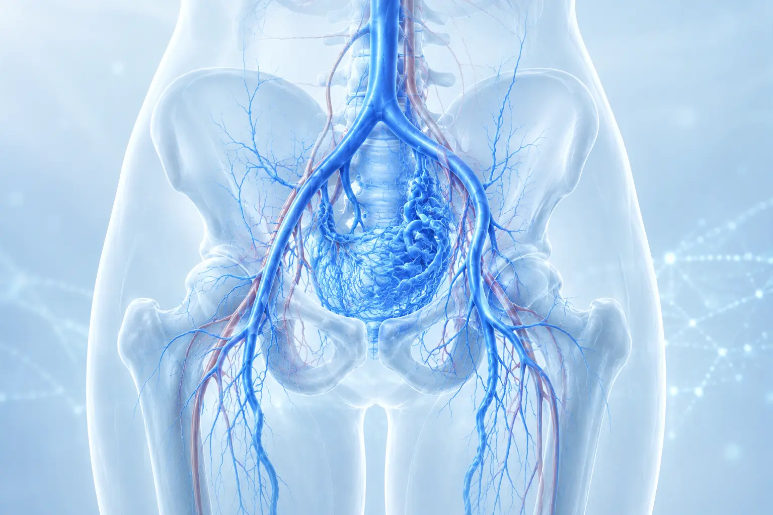
Understanding complex venous disease
Complex venous disease can result from obstruction, previous thrombosis, damaged valves or abnormal pelvic venous pathways. Because similar symptoms may have different causes, treatment begins by matching the patient’s history and examination with precise venous imaging.
Conditions assessed
After a deep vein thrombosis, residual obstruction or damage to venous valves can lead to persistent swelling, heaviness, pain, skin changes or venous ulcers.
Narrowing or blockage of the major veins in the pelvis can impair blood return from one or both legs. Causes include previous thrombosis, scarring or external compression.
In this anatomical compression pattern, the left iliac vein is compressed in the pelvis. It may cause no symptoms, or contribute to left-leg swelling, discomfort and deep vein thrombosis.
Abnormal reflux or obstruction in pelvic veins may cause chronic pelvic heaviness or pain and atypical varicose veins around the pelvis, groin or upper thigh.
A clot in a deep vein requires timely diagnosis. Treatment aims to prevent clot extension and pulmonary embolism while reducing the risk of long-term venous damage.
Narrowing or thrombosis of veins in the chest, shoulder or arm can cause swelling, visible collateral veins and discomfort, particularly after previous venous catheters or devices.
Advanced venous assessment
An anatomical finding alone does not always explain a patient’s symptoms. Clinical relevance is established by combining history, examination and appropriate imaging.
The symptom pattern and venous history are reviewed carefully.
Ultrasound evaluates deep and superficial vein patency, reflux, residual thrombosis and blood-flow patterns without radiation.
CT or MR venography may define pelvic and central venous anatomy, obstruction, collateral pathways and surrounding structures.
For selected patients being considered for intervention, catheter venography and intravascular ultrasound may provide detailed information from inside the vein.
Conservative management
These measures may reduce symptoms and support long-term venous health. They do not replace urgent assessment for a suspected blood clot or specialist evaluation of persistent symptoms.
Regular walking and calf-muscle activity
Avoiding prolonged immobility when possible
Individualized medical compression
Leg elevation for swelling when appropriate
Weight management and smoking cessation
Skin care and early treatment of venous wounds
Your treatment journey
Symptoms, previous thrombosis, interventions, medication and personal priorities are reviewed in detail.
Duplex ultrasound is often the first examination. CT or MR venography may be added when pelvic or central veins require assessment.
The clinical findings and imaging are considered together. Treatment is recommended only when the likely benefit justifies the risks.
Symptoms, access sites and any treated vein or stent are reviewed. Anticoagulation, compression and surveillance are individualized.
Frequently asked questions
Recommendations depend on symptoms, previous thrombosis, medical risks and the findings of targeted imaging.
Complex venous disease usually involves deep, pelvic or central veins, previous thrombosis, unusual venous anatomy, recurrent symptoms or several connected causes that require advanced imaging and individualized planning.
Duplex ultrasound is the first-line test for many venous problems. CT venography, MR venography, catheter venography or intravascular ultrasound may be needed when the pelvic or central veins cannot be assessed fully from the outside.
No. Anatomical compression can exist without causing disease. Intervention is considered only when symptoms, clinical findings and imaging support a clinically significant obstruction and the expected benefit outweighs the risks.
Yes. Some patients develop post-thrombotic syndrome because of persistent obstruction or valve damage. Symptoms may include swelling, heaviness, pain, skin changes and, in advanced cases, venous ulceration.
Yes. Pelvic reflux or obstruction can contribute to atypical or recurrent varicose veins around the pelvis, groin or upper thigh. The source should be identified before treatment is planned.
Anticoagulation is the main treatment for most patients. A smaller group with extensive, recent thrombosis and severe symptoms may be assessed for an additional catheter-based procedure.
Follow-up depends on the diagnosis and treatment. Patients with recurrent thrombosis, a venous stent, ongoing anticoagulation or chronic venous symptoms may require structured surveillance.
When to seek specialist advice
The information on this page is intended for general patient education and does not replace an individual medical examination, diagnosis or treatment recommendation. Procedure suitability and availability depend on the clinical indication and treating hospital.